


I spent many hours watching the World Cup over the summer. It was fascinating to learn the soccer balls are no longer stitched together. Why? Because stitches create spaces for leakage of water in and air out, which results in an imperfect shape and performance. The panels are instead fused with heat, creating a more perfect shape that is impermeable to leaking.
When fitting scleral lenses, measuring the ocular shape has been an evolving process. From using early anterior segment optical coherence tomography imaging to plot out angles of the cornea and sclera, we learned the nasal scleral angle was often smaller than the temporal scleral angle (Kojima et al, 2013; Ritzmann et al, 2018; Fadel, 2018). This asymmetry creates common barriers to getting a perfectly centered scleral lens and can lead to temporal and inferior decentration (Ritzmann et al, 2018; Fadel, 2018; DeNaeyer, 2020; Bergmann et al, 2023)
As modern technology has advanced, we have seen dramatic improvements in scanning capabilities, allowing for more consistent measurements. Many current devices rely on taking multiple images and stitching them together. While these methods are excellent improvements over historical options, they still create challenges of increased time for obtaining good scans and they open the door for errors to occur in lenses designed from them.
Case Description
A 34-year-old White male presented for a contact lens fitting. His spectacle correction was –3.25 –0.50 x 174 OD and –2.50 sphere OS. He had a history of giant papillary conjunctivitis, resulting in a relative intolerance to soft contact lens wear. He was unsatisfied from the movement of corneal GP lenses during sports and did not achieve satisfactory results with hybrids. He was also unsuccessful with 4 different scleral lens designs, adjusted at multiple offices. Upon discussing options, he stated that orthokeratology worked “OK” for him, but he experienced nighttime glare and preferred the sharpness of vision a scleral contact lens provided. No other ocular or systemic health issues were noted.
Discussion
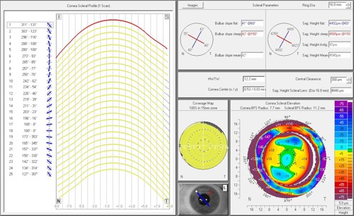
This patient had what many would call a “normal” eye, yet he wasn’t able to achieve success with multiple contact lens options. We proceeded to do a comprehensive tomography scan of each eye and came to understand the patient had a large corneal diameter, deep sag, and very asymmetric angles at a 16.00 mm chord, most notably found when comparing 48° on the inferior-temporal bulbar slope to 35° in the superior-nasal sector.
The single scan of each eye took less than 5 minutes from setup to completion and did not require image stitching. Historical technologies require multiple high-quality scans, followed by image stitching. Using the CSP Report Pro (OCULUS), we were able to design a lens from the data that matched the unique shape/angles of the eyes (Figure 1). The patient wore his new scleral lenses without fogging, lens awareness, or a tired feeling; all of which he previously struggled with in other scleral lens fitting processes.
Conclusion
Just like stitches in a soccer ball provide a space for leaks, stitching images of the eye together adds valuable chair time and creates challenges that can ultimately result in imperfect data. A single scan can consistently provide enormous data without the added time for multiple scans and image stitching, leading us one step closer to the goal of getting perfect measurements.
REFERENCES
1 Kojima R, Caroline PJ, Graff T, et al. Eye shape and scleral lenses. Contact Lens Spectr. 2013;28:38-43.
2 Ritzmann M, Caroline PJ, Börret R, Korszen E. An analysis of anterior scleral shape and its role in the design and fitting of scleral contact lenses. Contact Lens Anterior Eye. 2018;41(2):205-213. doi:10.1016/j.clae.2017.10.010
3 Fadel D. The influence of limbal and scleral shape on scleral lens design. Contact Lens Anterior Eye. 2018;41(4):321-328. doi:10.1016/j.clae.2018.02.003
4 DeNaeyer G. Scleral shape and its application for scleral lens fitting. Contact Lens Spectr. 2020;35:32-34, 36-37, 51.
5 Bergmann B, Wolffsohn JS, Bandlitz S. Scheimpflug imaging for grading and measurement of corneo-scleral-profile in different quadrants. Contact Lens Anterior Eye. 2023;46(2):101753. doi:10.1016/j.clae.2022.101753

To watch Dr. Rosen discuss this case, click here.