


This is a case in which a progressive myopia patient presented as an ideal candidate for orthokeratology (ortho-k). Using corneal topography, an initial lens was generated and ordered via a cloud-based platform. Guided design adjustments successfully refined the lens fit to improve treatment uniformity and achieve an optimal outcome.
Case Description
A 9-year-old white male presented for management of myopia. Previous records documented a myopic shift over the preceding 2 years. One year earlier, the left eye spectacle prescription measured –1.00 –1.25 x 078. At presentation, manifest refraction was –1.75 –1.50 x 082, representing an increase in both spherical and cylindrical refractive error.
Baseline corneal topography of the left eye measured 45.40 D @ 030° / 45.60 D @ 120°, demonstrating minimal corneal toricity. The anterior segment examination was unremarkable, with no accommodative or binocular vision abnormalities. Given the documented changes and otherwise healthy ocular findings, the patient was considered an appropriate candidate for ortho-k myopia management.
Corneal topography was uploaded into the Arise cloud platform (Bausch + Lomb), where the initial lens design was automatically generated. After review, no modifications were made, and the recommended left lens was ordered.
Following 1 night of wear, a slit lamp examination revealed a healthy anterior segment without corneal staining or adverse response. Manifest refraction improved to –0.50 –0.50 x 067, demonstrating a favorable initial response. The patient was instructed to continue nightly wear and return in 1 week.
At the 1-week follow-up, manifest refraction measured +1.00 –2.00 x 066, indicating overcorrection with induced against-the-rule astigmatism. Corneal health remained excellent. Follow-up topography was uploaded into the fitting system to evaluate management performance and determine whether lens modifications were needed.
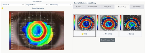
Topographic difference mapping demonstrated reasonable centration with a well-formed reverse curve, but a nonuniform treatment zone that was incomplete nasally with increased treatment effect inferotemporally. These findings correlated with the refractive outcome. Comparing the map to the built-in treatment pattern library, the fit was a mild “frowny face” consistent with subtle inferior lens decentration. After lens position and refractive findings were entered, the system recommended increasing the eccentricity of the alignment curves, effectively flattening the alignment curves to reduce sagittal depth and improve lens positioning.
The revised lens was dispensed, and after 1 week of overnight wear, manifest refraction improved to +0.25 –0.50 x 061. Corneal topography demonstrated a more uniform treatment zone with improved distribution of corneal flattening, consistent with the intended effect of the alignment curve modification. Corneal health remained uncompromised throughout treatment.
Discussion
Ortho-k outcomes do not always follow textbook expectations, making careful interpretation of corneal topography essential. Although the initial lens appeared reasonably centered, the system helped recognize a subtle pattern of inferior decentration and recommended a conservative alignment curve modification rather than changes to the base curve, reverse curve, or diameter. This single adjustment produced improved centration, a more uniform treatment zone, and a markedly better refractive outcome.
Conclusion
While fitting platforms provide evidence-based recommendations and built-in safety checks, they ideally complement rather than replace clinical judgment. When additional guidance is needed, clinicians can request consultant review directly within the platform. By simplifying lens design while maintaining practitioner oversight, modern ortho-k software can improve both clinical efficiency and first-fit success for clinicians at any level of experience.

To watch Dr. Skoner discuss this case, click here.