


While standard scleral lenses work beautifully for a broad range of patients, impression-based designs are typically reserved for complex, highly irregular eyes where standard fitting methods fail to achieve stability, comfort, or adequate visual acuity.
Case Description
A 76-year-old female was referred for therapeutic scleral lens evaluation due to persistent ocular surface symptoms. Her history included keratoconjunctivitis sicca associated with Sjögren’s syndrome, severe meibomian gland dysfunction (MGD), chronic trichiasis, severe punctate keratitis, and advanced primary open-angle glaucoma in both eyes, with a glaucoma shunt OS.
Her symptoms affected nearly every part of her day. She struggled at distance, intermediate, and near. The left eye felt constantly uncomfortable, while the right eye was less painful but remained blurred. Since tube shunt surgery OS, she had also noticed intermittent diplopia. She was significantly light sensitive and had undergone multiple eyelash cauterization procedures.
Her regimen included topical fluorometholone, 50% autologous serum tears, cyclosporine ophthalmic solution, tafluprost, latanoprost, brimonidine, nighttime lubricating ointment, oral doxycycline, varenicline nasal spray, hypochlorous acid lid spray, lid hygiene, and intermittent warm compresses. Despite this, she used artificial tears as often as every 15 minutes.
Best-corrected spectacle acuity was 20/40 OD and 20/200 OS. Slit lamp examination showed upper and lower lid trichiasis in both eyes, lid margin telangiectasia, blepharitis, severe meibomian gland atrophy, extensive lissamine green staining, and superficial punctate keratitis (SPK) grade 2 OD and 3+ OS.
A 17.0 mm diagnostic scleral lens was placed in both eyes, improving acuity to 20/25 OD and 20/25 OS. However, even with a relatively aligned lens, she reported significant edge awareness. Given her ocular surface disease (OSD), need for maximal protection, and importance of aligning over the glaucoma bleb and tube OS, we proceeded with an EyePrintPRO (by WAVE Eye Care) impression-based design. The goal was approximately 300 µm of clearance between the tube and posterior lens surface.
The initial lenses were designed at 20 mm OU to maximize therapeutic coverage. At dispense, the right lens was manageable, but she had significant difficulty applying and removing the left lens, even with a stand. Her advanced glaucoma and reduced visual field made accurate placement of a large round lens impractical.
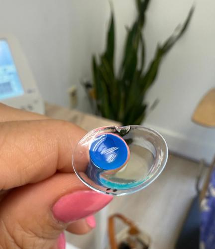
The left lens was redesigned as an ovate lens, measuring 20 mm horizontally and 16 mm vertically (Figure 1). This preserved horizontal coverage while making application more achievable.
She was also fit with an aberrometer-guided extended depth of focus multifocal, with counseling that advanced glaucomatous visual field loss, especially OS, could limit near function and adaptation.
After approximately 4 months of wear, her ocular surface and quality of life improved dramatically. She was able to drive independently again. She no longer needed artificial tears every 15 minutes. Corneal staining improved to grade 1 SPK in both eyes. Her regimen otherwise remained largely unchanged, with autologous serum and viscous gel placed into the lens bowl before application. She was also referred for intense pulsed light therapy and thermal pulsation treatment to further address severe MGD. Final acuity with the multifocal design was 20/20 OD, 20/25 OS, and 20/20 OU at distance, with approximately 20/50 near acuity.
Discussion
This case reinforced that scleral lens success is not defined by clearance, centration, or acuity alone. The first impression-based design provided ocular surface coverage, but the lens still had to work with the patient’s daily handling needs. For this patient, changing to an ovate design was the difference between a technically appropriate lens and one she could actually use.
Conclusion
In complex therapeutic cases, the final design must protect the ocular surface, respect surgical anatomy, and match the patient’s functional abilities. When those goals are met, scleral lenses can do more than improve the cornea, they can restore independence.
To watch Dr. Cerenzie discuss this case, click here.