


Scleral lenses are a powerful tool for vision rehabilitation in patients who have keratoconus and corneal transplants. However, poorly designed scleral lenses that fail to account for the true shape of the ocular surface can cause conjunctival compression, chronic inflammation, reduced vision, and, as this case illustrates, a granulomatous lesion.
Case Description
A 45-year-old male was referred for a specialty contact lens fitting. He had keratoconus in both eyes and a penetrating keratoplasty (PKP) in his right eye. With his habitual scleral lenses, he complained of constant redness and irritation, and saw 20/20-1 OD and 20/50-1 OS.
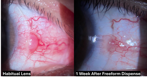
A 2.0 mm x 3.0 mm pyogenic granuloma on the nasal conjunctiva of his right eye was suspected to be secondary to scleral lens chafing. No improvement was noted with prednisolone acetate 1% BID, and the referring ophthalmologist suspected further treatment with a polidocanol 1% injection would be necessary.
The right eye examination confirmed the PKP with corneal neovascularization in the graft margin and a pyogenic granuloma with 2-3+ injection in the surrounding conjunctiva. The left eye demonstrated mild peripheral corneal neovascularization, a few Vogt’s striae, and mild apical scarring consistent with keratoconus.
With the patient’s habitual scleral lenses, the landing zone of both eyes demonstrated generalized trace to 2+ conjunctival blanching with 2-3+ injection near the pyogenic granuloma in the right eye. Prominent conjunctival impression rings were present upon removal of both lenses.
Scleral profilometry was performed, and Latitude (Visionary Optics) freeform lenses were ordered to follow the individualized shape of the scleral surface. The right lens was significantly vaulted to minimize the compressive force over the pyogenic granuloma.
At the first dispense, the customized landing zones appeared to significantly reduce compression in both eyes. The patient achieved 20/20 OD and 20/40+2 OS with small over-refractions in both eyes. One week later, the patient reported wearing his new lenses for 16 hours per day with good comfort. The pyogenic granuloma had diminished significantly, reducing central clearance from 305 µm to 189 µm.
Over the following few months, the right eye required 3 shape adjustments, as the shrinking granuloma left excessive clearance beneath the precision lift originally designed to vault it. Additionally, peripheral curves were progressively loosened to reduce landing zone compression as both eyes returned to their natural shape.
By the fourth pair of lenses, the pyogenic granuloma had completely resolved, revealing a mild pinguecula. The patient achieved 20/20 OD and 20/20-1 OS with good comfort.
Discussion
This case illustrates a complication that can arise when a spherically symmetric scleral lens is fitted on an eye with scleral asymmetry. A standard scleral lens landed on a preexisting pinguecula, creating chronic mechanical irritation and a pyogenic granuloma. The granuloma, in turn, further distorted the scleral shape and perpetuated the inflammatory cycle.
Switching to a profilometry-guided freeform design broke this cycle by evenly distributing the landing-zone load across the sclera’s actual topography.
This case also demonstrates that freeform fitting is not a one-time event when the underlying ocular surface is actively changing. As the granuloma resolved and the eye returned to its natural shape, sequential lens modifications were required.
Conclusion
In the end, this patient achieved complete resolution of his pyogenic granuloma, 20/20 vision in both eyes, and was wearing his scleral lenses comfortably for 16 hours per day. Profilometry-guided scleral lens design made this outcome possible by matching the lens to the patient’s eye rather than asking the eye to accommodate the lens.

To watch Dr. Shin discuss this case, click here.