


Exposure keratopathy results from incomplete eyelid closure and tear film instability, leading to corneal desiccation, epithelial breakdown, and symptoms of irritation, pain, and fluctuating vision (Rodriguez-Garcia et al, 2025). Management strategies range from lubrication and eyelid taping to surgical intervention, depending on severity (Rodriguez-Garcia et al, 2025). Scleral lenses provide an alternative by maintaining a continuous protective fluid reservoir over the cornea, promoting surface healing and improving patient comfort (Rodriguez-Garcia et al, 2025; Harthan and Shorter, 2018)
Case History
A 59-year-old woman was referred for scleral lens evaluation for persistent exposure keratopathy OD secondary to chronic facial nerve palsy. Her history was notable for a prior pontine stroke as well as long-standing Bell’s palsy with residual right facial weakness and lagophthalmos. She reported daily irritation, tearing, and fluctuating blur that worsened throughout the day.
Prior management included artificial tears, nighttime ointment, and intermittent lid taping. She had also undergone oculoplastic reconstruction, including midface lift, canthoplasty, and lid repositioning procedures, followed by ectropion repair, with incomplete resolution of symptoms.
Best-corrected visual acuity (BCVA) was approximately 20/25 OD and 20/20 OS. Slit lamp examination OD revealed 1 mm to 2 mm lagophthalmos with intact Bell's reflex, inferior punctate epithelial erosions, and reduced tear breakup time.
Given persistent surface disease despite medical and surgical management, a scleral lens was recommended to provide continuous daytime corneal protection.
The patient was initially fit with a SynergEyes VS scleral lens (CooperVision) OD and a daily disposable soft lens OS. The initial fit demonstrated good central clearance, mild edge awareness, and variable fogging during wear.
Refinements were made to improve alignment and comfort. Lens diameter was increased from 16.0 mm to 17.5 mm, and the scleral landing zone was steepened to optimize scleral alignment and reduce edge awareness. Evaluation in secondary gaze informed additional adjustments to improve peripheral alignment. These combined changes resulted in improved lens centration, stable limbal clearance, and enhanced comfort throughout the day.
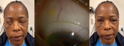
Discussion
The patient achieved visual acuity of approximately 20/20 OD with improved comfort and daily function. Corneal integrity improved, with reduction of epithelial compromise and more stable ocular surface findings.
This case demonstrates the effectiveness of scleral lenses in managing paralytic exposure keratopathy. A structured, iterative approach to lens design and surface management can successfully optimize both fit and function. Scleral lenses provide sustained ocular surface protection and represent a valuable therapeutic option for patients who have chronic exposure.
Conclusion
With expanding availability and awareness of scleral lens therapy, earlier intervention is increasingly possible for patients who have paralytic lagophthalmos. Timely consideration of scleral lenses may reduce prolonged exposure-related corneal disease and improve patient comfort, particularly in complex cases managed across subspecialties.
REFERENCES
1 Rodriguez-Garcia A, Ruiz-Lozano RE, Barcelo-Canton RH, Marines-Sanchez HM, Homar Paez-Garza J. The etiologic and pathogenic spectrum of exposure keratopathy: diagnostic and therapeutic implications. Surv Ophthalmol. 2025;70(5):882-899. doi:10.1016/j.survophthal.2025.03.001
2 Scanzera AC, Ahmad A, Shorter E. Adjunct use of therapeutic scleral lens for exposure keratopathy after severe chemical burn. Case Rep Ophthalmol. 2021;12(1):243-247. doi:10.1159/000511223
3 Harthan JS, Shorter E. Therapeutic uses of scleral contact lenses for ocular surface disease: patient selection and special considerations. Clin Optom (Auckl). 2018;10:65-74. doi:10.2147/OPTO.S144357

To watch Dr. Scanzera discuss this case, click here.