


The use of scleral contact lenses has been a rapidly growing aspect of eye care, and it is commonly used to manage corneal ectasias, ocular surface disease, and a variety of refractive errors (Harthan and Shorter, 2018). A less common, but very important utilization of scleral lenses is for the visual correction of posttraumatic corneal conditions. These patients often find themselves with high amounts of induced higher-order aberrations that cannot be corrected with more traditional visual correction, which can negatively affect overall quality of life. This case report highlights a successful fitting and restoration of visual function for a patient following penetrating corneal trauma.
Case Description
A 38-year-old Hispanic male presented with vision loss in his left eye following penetrating corneal trauma that occurred 3 months prior. He reported having seen an eyecare provider immediately following the trauma, who had successfully managed injury but told him that he would have continued vision loss in that eye due to corneal scarring. The patient desired a second opinion on his options for visual improvement.
On evaluation, his best-corrected vision for his right eye was 20/20 with a –0.50 DS and 20/80 with –2.50 –2.50 x 002 spectacle prescription.
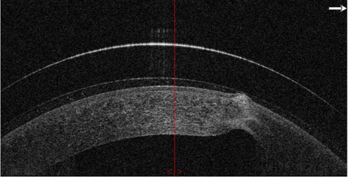
Slit lamp examination revealed a 3 mm long scar that translated through the entire thickness of the cornea and was adjacent to the visual axis. Axial pellucid ring corneal topography of the left eye revealed irregular steepening corresponding with the location of the corneal scar. Total higher-order aberrations demonstrated a significant decrease in visual quality with traditional spectacle correction. Despite the low prescription in the right eye, it was decided to fit both eyes. Due to the central location of the corneal scarring, the InSight Prolate SAG 4.500 scleral lens (Metro Optics) was chosen for both eyes.
Although the scar was only present in the left eye, the overall profile of each eye was not significantly different, so it was determined a scleral lens with the same sagittal height would be appropriate for both eyes.
With the initial fitting lens of the left eye, minimal central clearance was noted (approximately 100 µm) centrally, and light corneal touch was noted over the scar. There was good limbal coverage and a good landing zone with no compression or impingement. A good overall fit was achieved with the trial lens in the right eye. To improve clearance, the sagittal height was increased by 150 µm and the lens was ordered.
With the over-refraction, 20/20 vision was restored and the patient noted significant improvement in depth perception.
Discussion
Managing severe corneal trauma can be very complicated, and is often determined “complete” when the wound is sealed and any infection is treated/prevented. However, part of the management of these patients should include the restoration of visual function, to the extent possible.
Conclusion
Scleral lenses are a great resource for correcting higher-order aberrations induced by traumatic alterations to the ocular surface, even with the presence of significant corneal scarring. Wavefront-guided over-refractions and their implementation into scleral lenses have also further advanced our ability to improve the quality of life of these patients.
REFERENCE
1 Harthan JS, Shorter E. Therapeutic uses of scleral contact lenses for ocular surface disease: patient selection and special considerations. Clin Optom (Auckl). 2018;10:65-74. doi:10.2147/OPTO.S144357

To watch Dr. Kemp discuss this case, click here.